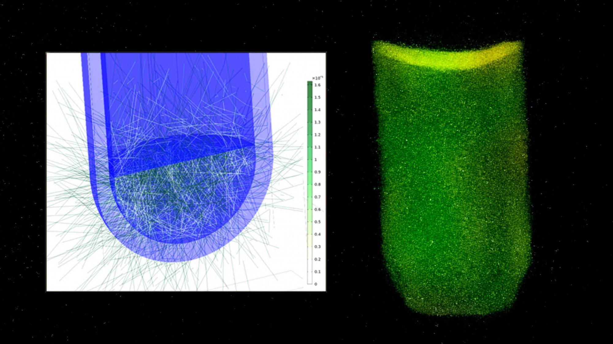
Jatkuvatoiminen ATP-perusteinen mikrobitasomääritys prosessiteollisuuteen
ATP
Rahoittajat


Projektin tiedot
Projektin kesto
-
Rahoittaja
Euroopan rakenne- ja investointirahastot - Euroopan aluekehitysrahasto (EAKR)
Rahoituksen määrä
440 556 EUR
Projektin koordinaattori
Oulun yliopisto
Yhteystiedot
Yhteyshenkilö
Projektin kuvaus
ATP-hanke keskittyy teollisuudessa olevien prosessien mikrobitason määritykseen mikrobiologian ja mittaustekniikan keinoin.
Hankkeen kehittämisen kohteena ovat vesi-intensiivisten teollisuusprosessien parempi kontrolloitavuus, materian ja energian käytön optimointi sekä pienentää ympäristökuormitusta.
Hankeen tavoitteena on parantaa teollisuussektorien toiminta edellytyksiä Kainuussa ja mahdollistaa uusia investointeja. Teollisuusprosessien tarpeisiin hankkeessa selvitetään teolliseen käyttöön skaalautuvaa mittausjärjestelmää.
Hankkeen painopistealueen kohteena ovat metsäbiotalous, vedenpuhdistus ja kaivannaisala.
Tavoitteena on saada selville mikrobimäärän tunnistamisratkaisun sovellettavuus erilaisiin teollisuusprosesseihin.
Kainuun teollisuussektorin tarpeisiin hankkeen tavoitteena on tuottaa tietoa mittalaite- ja järjestelmävalintoihin.
Hankkeessa tunnistettavien ratkaisujen ympärille kootaan jatkokehitystä varten soveltuva ekosysteemi.
Hankkeen toteuttaa Oulun yliopiston Mittaustekniikan yksikkö MITY Kajaanissa. Hankkeen kokonaiskustannusarvio on 440k euroa.
Projektin tulokset
https://www.procedia-esem.eu/pdf/issues/2025/no4/115_Raykhel_25.pdf
Kirjallisuusselvityksen viitteitä:
[1] Bhuyan S., Yadav M., Giri S., Begum S., Das S., Phukan A., Priyadarshani P., Sarkar S., Jayswal A., Kabyashree K., Kumar A., Mandal M., Ray S., (2023) Microliter spotting and micro-colony observation: A rapid and simple approach for counting bacterial colony forming units, Journal of Microbiological Methods, 207, 106707.
[2] Daims H., Wagner M. (2007) Quantification of uncultured microorganisms by fluorescence microscopy and digital image analysis. Appl Microbiol Biotechnol 75. 237–248.
[3] Brown M., Hands C., Coello-Garcia T., Sani B., Ott A., Smith S., Davenport R., (2019) A flow cytometry method for bacterial quantification and biomass estimates in activated sludge, Journal of Microbiological Methods,160. 73-83.
[4] Lomakina G., Modestova Y., Ugarova N. (2015) Bioluminescence assay for cell viability. Biochemistry (Mosc). 80(6).
[5] Lundin A. (2014) Optimization of the firefly luciferase reaction for analytical purposes. Adv Biochem Eng Biotechnol. 145. 31-62.
[6] McElroy W. (1947) The Energy Source for Bioluminescence in an Isolated System. Proc Natl Acad Sci U S A. 33(11). 342-5.
[7] Marques S., Esteves da Silva J. (2009) Firefly bioluminescence. A mechanistic approach of luciferase catalyzed reactions, IUBMB Life. 61. 6–17.
[8] Inouye S. (2010) Firefly luciferase: an adenyl-ate-forming enzyme for multicatalytic functions. Cell. Mol. Life Sci. 67. 387–404.
[9] Leitão J., Esteves da Silva J. (2010) Firefly luciferase inhibition. J Photochem Photobiol B. 101(1). 1-8.
[10] Berthold F., Tarkkanen V. (2013) Luminometer development in the last four decades: recol-lections of two entrepreneurs. Luminescence. 28(1). 1-6.
[11] Lundin A. (2000) Use of firefly luciferase in ATP-related assays of biomass, enzymes, and metabolites. Methods Enzymol. 305. 346-70.
[12] Gerba C., Pepper I. (2019) Environmental and Pollution Science (3rd Edition).
[13] Yaginuma H., Kawai S., Tabata K., Tomiyama K., Kakizuka A., Komatsuzaki T., Noji H., Imamura H. (2014) Diversity in ATP concentrations in a single bacterial cell population revealed by quantitative single-cell imaging. Sci Rep.4. 6522.
[14] Ren L., Qi Y., Cao F. (2025) Study on the framework of ATP energy cycle system in Escherichia coli. Appl Microbiol Biotechnol. 109, 42.
[15] Taylor S., Winter S. (2020) Salmonella finds a way: Metabolic versatility of Salmonella enterica serovar Typhimurium in diverse host environments. PLoS Pathog. 16(6): e1008540.
[16] van der Kooij D., Bakker G., Italiaander R., Veenendaal H., Wullings B. (2017) Biofilm Composition and Threshold Concentration for Growth of Legionella pneumophila on Surfaces Exposed to Flowing Warm Tap Water without Disinfectant. Appl Environ Microbiol. 83(5). e02737-16.
[17] Ghaffar N., Connerton P., Connerton I. (2015) Filamentation of Campylobacter in broth cultures. Front Microbiol. 6. 657.
[18] Xu J., Abe K., Kodama T., Sultana M., Chac D., Markiewicz S., Matsunami H., Kuba E., Tsunoda S., Alam M., Weil A., Nakamura S., Yamashiro T. (2025) The role of morphological adaptability in Vibrio cholerae's motility. mBio. 16(1). e0246924.
[19] Khan S., Zhang X., Witola W. (2022) Cryptosporidium parvum Pyruvate Kinase Inhibitors With in vivo Anti-cryptosporidial Efficacy. Front Microbiol. 12. 800293.
[20] Braissant O., Astasov-Frauenhoffer M., Waltimo T., Bonkat G. (2020) A Review of Methods to Determine Viability, Vitality, and Metabolic Rates in Microbiology. Front Microbiol. 11. 547458.
[21] Bajerski F., Stock J., Hanf B., Darienko T., Heine-Dobbernack E., Lorenz M., Naujox L., Keller E., Schumacher H., Friedl T., Eberth S., Mock H., Kniemeyer O., Overmann J. (2018) ATP Content and Cell Viability as Indicators for Cryostress Across the Diversity of Life. Front Physiol. 9. 921.
[22] Appanna V., Auger C., Thomas S., Omri A. (2014) Fumarate metabolism and ATP production in Pseudomonas fluorescens exposed to nitrosative stress. Antonie Van Leeuwenhoek. 106(3). 431-8.
[23] Li B., Chen X., Yang J-Y., Gao S., Bai F. (2024) Intracellular ATP concentration is a key regulator of bacterial cell fate. J Bacteriol. 206(12). e0020824.
[24] Qiu X., Hu X., Tang X., Huang C., Jian H., Lin D. (2024) Metabolic adaptations of Macrobacterium sediments YLB-01 in deep-sea high-pressure environments. Appl Microbiol Biotech-nol. 108(1). 170.
[25] Pakostova E., Mandl M., Omesova Pokorna B., Diviskova E., Lojek A. (2013) Cellular ATP Changes in Acidithiobacillus ferrooxidans Cul-tures Oxidizing Ferrous Iron and Elemental Sulfur. Geomicrobiology Journal, 30,1, 1-7.
[26] Camacho D., Frazao R., Fouillen A., Nanci A., Lang B., Apte S., Baron C., Warren L. (2020) New Insights Into Acidithiobacillus thiooxidans Sulfur Metabolism Through Coupled Gene Expression, Solution Chemistry, Microscopy, and Spectroscopy Analyses. Front Microbiol. 11. 411.
[27] Redsven I., Kymalainen H., Pesonen-Leinonen E., Kuisma R., Ojala-Paloposki T., Hautala M., Sjoberg A. (2007) Evaluation of a bioluminescence method, contact angle measurements and topography for testing the cleanability of plastic surfaces under laboratory conditions. Appl. Surf. Sci. 253, 5536–5543.
[28] Uzlova E., Zimatkin S. (2021) Cellular ATP Synthase. Biol Bull Rev. 11, 134–142.
[29] Okanojo M., Miyashita N., Tazaki A., Tada H., Hamazoto F., Hisamatsu M., Noda H. (2017) Attomol-level ATP bioluminometer for detecting single bacterium. Luminescence. 32(5):751-756.
[30] Romanova N., Brovko L., Ugarova N. (1997) Comparative assessment of methods of intracellular ATP extraction from different types of microorganisms for bioluminescent determination of microbial cells, Appl. Biochem. Microbiol. 33, 306-311.
[31] Efremenko E., Senko O., Stepanov N, Maslova O., Lomakina G., Ugarova N. (2022) Review Luminescent Analysis of ATP: Modern Objects and Processes for Sensing. Chemosen-sors. 10, 493.
[32] Thorne N., Inglese J., Auld D. (2010) Illuminating insights into firefly luciferase and other bioluminescent reporters used in chemical biology. Chem Biol. 17(6):646-57.
[33] Shama G., Malik D. (2013) The uses and abuses of rapid bioluminescence-based ATP as-says. Int J Hyg Environ Health. 216(2). 115-25.
[34] Tetro J., Sattar S. (2021) The Application of ATP Bioluminescence for Rapid Monitoring of Microbiological Contamination on Environmental Surfaces: A Critical Review, InfectionControl.tips.
[35] Bochdansky A., Stouffer A., Washington N. (2021) Adenosine triphosphate (ATP) as a metric of microbial biomass in aquatic systems: new simplified protocols, laboratory validation, and a reflection on data from the literature. Limnol Oceanogr Methods, 19: 115-131.
[36] Ratnawulan R., Fauzi A., Arma A. (2019) Effects of heavy metals on intensity and coefficient of inhibition of firefly bioluminescence, Journal of Physics Conference Series, 1185:012016.
[37] Abushaban A., Mangal M., Salinas-Rodriguez S., Nnebuo C., Mondal S., Goueli S., Schippers J., Kennedy M. (2017) Direct measurement of ATP in seawater and application of ATP to monitor bacterial growth potential in SWRO pre-treatment systems. Desalination and Water Treatment. 99. 91-101.
[38] Yawata S., Noda K., Shimomura A. (2021) Mutant firefly luciferase enzymes resistant to the inhibition by sodium chlo-ride. Biotechnol Lett 43, 1585–1594.
[39] Efremenko E., Senko O., Stepanov N., Mareev N., Volikov A., Perminova I. (2020) Suppression of Methane Generation during Methanogenesis by Chemically Modified Humic Compounds. Antioxidants. 9. 1140.
[40] Kotlobay A., Kaskova Z., Yampolsky I. (2020) Palette of Luciferases: Natural Biotools for New Applications in Biomedicine. Acta Naturae. 12(2). 15-27.
[41] Ismayilov I., Stepanov N., Efremenko E. (2015) Evaluation of biocidal properties of vegetable oil-based corrosion inhibitors using bioluminescent enzymatic method. Moscow Univ. Chem. Bull. 70, 197–201.
[42] Efremenko E., Ugarova N., Lomakina G., Senko O., Stepanov N., Maslova O., Aslanly A., Lyagin I. (2022) М.: Publishing House «SCIEN-TIFIC LIBRARY», 376.
[43] Hammes F., Goldschmidt F., Vital M., Wang Y., Egli T. (2010) Measurement and interpretation of microbial adenosine tri-phosphate (ATP) in aquatic environments. Water Res. 44(13). 3915-23.
[44] Siebel E., Wang Y., Egli T., Hammes F. (2008) Correlations between total cell concentration, total adenosine tri-phosphate concentration and heterotrophic plate counts during microbial monitoring of drinking water. Drink. Water Eng. Sci. Discuss. 1. 1–6.
[45] Magic-Knezev A., van der Kooij D. (2004) Optimisation and significance of ATP analysis for measuring active biomass in granular activated carbon filters used in water treatment, Water Res. 38. 3971–3979.
[46] Prest E., Hammes F., van Loosdrecht M., Vrouwenvelder J. (2016) Biological stability of drinking water: controlling factors, methods, and challenges, Front.Microbiol. 7. 45.
[47] Eydal H., Pedersen K. (2007) Use of an ATP assay to determine viable microbial biomass in Fennoscandian Shield groundwater from depths of 3–1000 m, J. Microbiol. Methods. 70. 363–373.
[48] Prest E., Weissbrodt D., Hammes F., van Loosdrecht M., Vrouwenvelder J. (2016) Long-Term Bacterial Dynamics in a Full-Scale Drinking Water Distribution System. PLoS One. 11(10). e0164445.
[49] Vital M., Dignum M., Magic-Knezev A., Ross P., Rietveld L., Hammes F. (2012) Flow cytometry and adenosine tri-phosphate analysis: alternative possibilities to evaluate major bacteriological changes in drinking water treatment and distribution systems. Water Res. 46(15). 4665-76.
[50] Hansen C., Kerrouche A., Tatari K., Rasmussen A., Ryan T., Summersgill P., Desmulliez M., Bridle H., Albrechtsen H. (2019) Monitoring of drinking water quality using automated ATP quantification. J Microbial Methods. 165. 105713.
[51] Lee Y., Imminger S., Czekalski N., von Gunten U., Hammes F. (2016) Inactivation efficiency of Escherichia coli and autochthonous bacteria during ozonation of municipal wastewater effluents quantified with flow cytome-try and adenosine tri-phosphate analyses. Water Res. 101. 617-627.
[52] Li G., Yu T., Wu Q., Lu Y, Hu H. (2017) Development of an ATP luminescence-based method for assimilable organic carbon determination in reclaimed water. Water Research. 123, 345-352.
[53] Amy G., Salinas Rodriguez S, Kennedy M., Schippers J., Rapenne S., Remize P., Barbe C., de O. Manes C., West N., Lebaron P., van der Kooij P., Veenendaal H, Schaule G., Petrowski K., Huber S., Sim L., Ye Y., Chen V., Fane A. (2011) Water quality assessment tools, Mem-brane-based Desalination: An Integrated Ap-proach (MEDINA), IWA Publishing, 3–32.
[54] Abushabana A., Salinas-Rodrigueza S., Mangala M., Mondalc S., Gouelic S., Knezeve A., Vrouwenvelderf J., Schippersa J., Kennedya M. (2019) ATP measurement in seawater reverse osmosis systems: Eliminating seawater matrix effects using a filtration-based method. Desalination. 453. 1-9,
[55] Slowik G., Richer R., Stryjska A., Dabrowski P. (2024) Bacteria Acidithiobacillus ferrooxidans, terrestrial analogue of extraterrestri-al microorganisms? International Journal of Astrobiology. 23. e22.
[56] Pakostova E., Mandl M., Tuovinen O. (2013) Cellular ATP and biomass of attached and planktonic sulfur-oxidizing Acidithiobacillus ferrooxidans. Process Biochemistry. 48(11). 1785-1788.
[57] Johnson D., Pakostova E. (2021) Dissolution of Manganese (IV) Oxide Mediated by Aci-dophilic Bacteria, and Demonstration That Manganese (IV) Can Act as Both a Direct and Indirect Electron Acceptor for Iron-Reducing Acidithiobacillus spp. Geomicrobiology Journal. 38:7. 570-576.
[58] Chen G., Shi H., Ding H., Zhang X., Gu T., Zhu M., Tan W. (2022) Multi-scale analysis of nickel ion tolerance mechanism for thermophilic Sulfobacillus thermosulfidooxidans in bioleaching. J Hazard Mater. 443(Pt B). 130245.
[59] Izquierdo-Fiallo K., Muñoz-Villagrán C., Schimpf C., Mardonez M., Rafaja D., Schlömann M., Tello M., Orellana O., Levicán G. (2025) Adaptive response of the holdase chaperone network of Acidithiobacillus ferrooxidans ATCC 23270 to stresses and energy sources. World J Microbiol Biotechnol. 41(4). 121.
[60] Salimiyan rizi K., Ashrafi A. (2023) Biosensors, mechatronics, & microfluidics for early detection & monitoring of microbial corrosion: A comprehensive critical review, Results in Materi-als, 18.100402, ISSN 2590-048X.
[61] Mentu J., Pirttijärvi T., Lindell H. Salkinoja-Salonen M. (1997) Microbiological control of pigments and fillers in paper industry. In The Fundamentals of Papermaking Materials, Trans. of the XIth Fund. Res. Symp. Cambridge, (C.F. Baker, ed.), 955–993, FRC, Manchester, 2018.
[62] Kiuru J., Tsitko I., Sievänen J., Wathen R. (2010). Biocide strategies for fine paper. BioResources. 5(2), 514-524.
[63] Lund L., Rice L., Luth E., Sirois D. (2012) The Use of On-line Monitoring Tools and Advanced Analytical Techniques to Optimize Bio-control Treatment Strategies for Papermaking Systems. DOI: 10.13140/2.1.5021.9523. Confer-ence: PaperCon 2012At: New Orleans
[64] Rice L., Lund L. (2013) Detection and quan-tification of nucleic acid to assess microbial biomass in paper defects and machine felts, United States Patent No US 8,613,837 B2.
[65] Flemming H., Meier M., Schild T. (2013) Mini-review: microbial problems in paper production. Biofouling. 29(6). 683-96.
[66] Zumsteg A., Urwyler S., Glaubitz J. (2017) Characterizing bacterial communities in paper production-troublemakers revealed. Microbiologyopen. 6(4). e00487.
[67] Morohoshi T., Oi T., Aiso H., Suzuki T., Okura T., Sato S. (2018) Biofilm Formation and Degradation of Commercially Available Biodegradable Plastic Films by Bacterial Consortiums in Freshwater Environments. Microbes Environ. 33(3). 332-335.
[68] Hosoe A., Suenaga T., Sugi T., Iizumi T., Nagai N., Terada A. (2020) Complete Genome Sequence of Pseudomonas putida Strain TS312, Harboring an HdtS-Type N-Acyl-Homoserine Lactone Synthase, Isolated from a Paper Mill. Microbiol Resour Announc. 9(13). e00055-20.
[69] Pathak P., Kumar V., Bhardwaj N., Sharma C. (2021) Slime control in paper mill using bio-logical agents as biocides. Physical Sciences Reviews. 6.
[70] Kaur A., Gautam L., Balda S., Capalash N., Sharma P. (2022) Triclosan controls pleiotropically the paper-deteriorating bacterial community in paper mill, International Biodeterioration & Biodegradation, 173. 105455.
[71] Ali A., Altemimi A., Alhelfi N., Ibrahim S. (2020) Application of Biosensors for Detection of Pathogenic Food Bacteria: A Review. Biosensors (Basel). 10(6):58.
[72] Rajapaksha P., Elbourne A., Gangadoo S., Brown R., Cozzolino D., Chapman J. (2019) A review of methods for the detection of pathogenic microorganisms. Analyst. 144(2). 396-411.